Transmitted Illumination
Transmitted Illumination: Shedding Light on the World of Microscopy
Microscopy is an essential tool for scientists and researchers, enabling them to zoom in on the infinitely small and explore the intricate world of biological structures. However, none of this would be possible without proper illumination, which is often overlooked despite being one of the most critical components of microscopy. In this article, we will explore transmitted illumination, one of the most commonly used methods, and its importance in achieving accurate results and interpreting data.
When it comes to transmitted illumination, several pain points frustrate researchers. One of the most common issues is obtaining consistent illumination across the field of view, resulting in uneven lighting and compromised image quality. Another issue is achieving the right intensity and wavelength to excite the sample without damaging it. Finally, light scattering and absorption can cause distortions, making it challenging to interpret results accurately. Addressing these issues requires an in-depth understanding of the principles of transmitted illumination.
The target of transmitted illumination is to pass light through a sample, allowing the observer to visualize its internal structure. This is achieved by placing the sample between the light source and the objective lens, with the intensity and angle of illumination adjusted to achieve optimal contrast. Transmitted illumination is commonly used in brightfield microscopy, phase contrast microscopy, and darkfield microscopy, each with its unique benefits and challenges.
In summary, transmitted illumination is a fundamental aspect of microscopy that requires careful consideration to achieve accurate results. It is essential to understand the principles underlying transmitted illumination to select the right method for each sample, optimize the lighting conditions, and minimize artifacts that can compromise the data. By doing so, researchers can unlock the full potential of microscopy to explore new frontiers in science and medicine.
Transmitted Illumination and Brightfield Microscopy
Brightfield microscopy is one of the most common methods used to observe transparent samples, making transmitted illumination a critical aspect of its success. When using transmitted illumination, the light passes through the sample, and the differences in refractive index cause the light to bend and create contrast. This method is highly sensitive to density variations, making it ideal for observing cells and other biological structures. However, achieving sufficient contrast and minimizing artifacts requires careful adjustment of the illuminator and objective lens.
When I first started working with brightfield microscopy, I struggled with getting consistent illumination across the field of view. It was frustrating to see my images with uneven lighting and hot spots that obscured the delicate structures I was trying to observe. However, by experimenting with different illumination angles and adjusting the diaphragm, I was able to achieve the results I desired. It was a valuable lesson in the importance of proper illumination and its impact on the quality of the results.
Transmitted Illumination and Phase Contrast Microscopy
Phase contrast microscopy is another popular method that utilizes transmitted illumination to visualize transparent samples. In this method, the light passes through a phase plate that causes a phase shift in the light, creating contrast between the sample and background. This method is ideal for observing live cells in real-time, as it does not require staining or fixation. However, it also requires careful alignment and calibration of the phase plate, which can be challenging for beginners.
When I first started using phase contrast microscopy, I found it challenging to obtain consistent phase contrast across the sample. It was frustrating to see artifacts and halos that made it challenging to interpret my data. However, by carefully adjusting the alignment and intensity of the illuminator, and using high-quality phase plates, I was able to overcome these challenges and obtain high-quality results. It was a valuable lesson in the importance of patience and attention to detail in microscopy.
Transmitted Illumination and Darkfield Microscopy
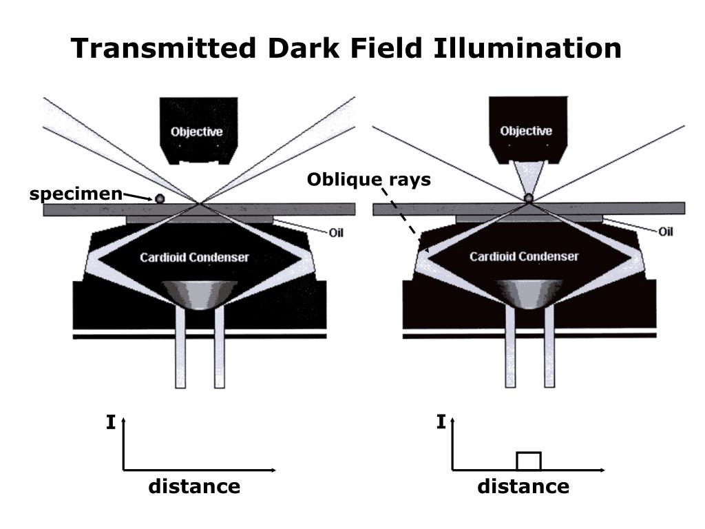
Darkfield microscopy is a unique method that utilizes transmitted illumination at oblique angles, making it ideal for observing highly refractive samples, such as bacteria and spores. In this method, only the scattered light is observed, creating a bright image against a dark background. This method is ideal for observing the structure and movement of transparent specimens that are invisible under other methods. However, it also requires specialized equipment and careful adjustment of the illuminator.
Darkfield microscopy is an exciting method that I used to observe the behavior of bacterial colonies. By adjusting the angle and direction of the illuminator, I was able to obtain detailed images of the colonies' structure and movement, providing valuable insights into their behavior and growth patterns. It was an excellent example of the power of transmitted illumination and its ability to reveal new insights and discoveries.
Transmitted Illumination and Fluorescence Microscopy
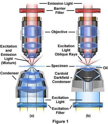
Fluorescence microscopy is a powerful method that utilizes transmitted illumination to excite fluorescent dyes and proteins, allowing them to emit light of different colors and intensity. This method is highly sensitive and specific, making it ideal for observing live cells and tissues in real-time. However, it also requires careful selection of the right light source and filter, as well as optimization of the excitation and emission wavelengths to achieve optimal results.
Small adjustments in the lighting conditions can have a significant impact on the results, making it essential to pay close attention to the illumination settings. When I first started using fluorescence microscopy, I struggled with obtaining the right balance between the excitation and emission, resulting in low signal-to-noise ratio and compromised data. However, by experimenting with different light sources and filters, and utilizing specialized software, I was able to enhance my results and obtain valuable insights into the biological structures I was observing.
Questions and Answers About Transmitted Illumination
1. Why is transmitted illumination critical for microscopy?
Transmitted illumination is essential for microscopy, as it enables researchers to visualize the internal structure of samples. By shining light through the sample, researchers can obtain information about its composition, density, and morphology, providing valuable insights into biological structures and processes.
2. What are the different types of transmitted illumination?
There are several types of transmitted illumination used in microscopy, including brightfield, phase contrast, darkfield, and fluorescence. Each type of illumination has its unique benefits and challenges, making it essential to understand the principles underlying each method to select the right one for each sample.
3. How can researchers optimize transmitted illumination for accurate results?
To optimize transmitted illumination, researchers should carefully adjust the intensity, angle, and wavelength of the light source to achieve optimal contrast and minimize artifacts. It is also essential to select the right method for each sample, depending on its composition, density, and morphology.
4. What are some common issues that researchers face when using transmitted illumination?
Some common issues that researchers face when using transmitted illumination include uneven illumination across the field of view, light scattering and absorption, and insufficient contrast. Addressing these issues may require adjusting the illuminator and objective lens, selecting the right method for each sample, and minimizing artifacts that can compromise the data.
Conclusion of Transmitted Illumination
Transmitted illumination is a critical aspect of microscopy that requires careful attention to achieve accurate results and interpret data. By understanding the principles underlying each method and optimizing the lighting conditions, researchers can unlock the full potential of microscopy to explore new frontiers in science and medicine. From brightfield to darkfield, phase contrast to fluorescence, transmitted illumination provides a window into the intricate world of biological structures, revealing new insights and discoveries that can transform our understanding of the world around us.
Gallery
Fluorescence Microscopy - Transmitted Light Illumination | Olympus LS

Photo Credit by: bing.com / microscope light illumination transmitted fluorescence microscopy cone olympus optical primer techniques tikz rays draw eye anatomy emission
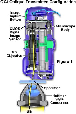
Molecular Expressions: Science, Optics And You - Intel Play QX3

Photo Credit by: bing.com / illumination oblique transmitted microscope optics
Transmitted Illumination | KEYENCE America

Photo Credit by: bing.com / illumination transmitted keyence
PPT - Optical Microscopy PowerPoint Presentation, Free Download - ID

Photo Credit by: bing.com / illumination transmitted field dark microscopy optical distance ppt powerpoint presentation specimen oblique rays slideserve
Transmitted Illumination | KEYENCE America

Photo Credit by: bing.com / transmitted illumination keyence